Chronic Arthritis in Knees: Symptoms, Causes and Management

Written by Julia Zhu - UPG Physiotherapist & Pilates Instructor

People reporting symptoms such as joint pain, stiffness and swelling, mainly in hands, knees and hips might be experiencing a chronic disease and the most common form of chronic arthritis known as Osteoarthritis (OA). Although there are no specific causes, the common risk factors for OA include joint injuries, excess weight, older age, female and repetitive joint-loading tasks. 

The knees are among the joints most affected by OA. It is characterised by degeneration of the knee’s articular cartilage. The damaged or missing knee cartilage can cause friction between bones and changes to bone tissue, which can cause pain. Knee OA also involves changes to the bone underneath the cartilage. 

Healthy Joint VS. Joint with Osteoarthritis - Source: AIHW 2015

Although currently there is no cure for OA, there are many non-surgical and non-drug treatments for people who suffer from this condition.

Regular exercise:

  • Strong evidence shows that land-based exercise such as muscle strengthening exercise, walking and Tai-chi is highly recommended. Other options include stationary bikes and Yoga. 

  • Dosage of strengthening training - Speak to your UPG physiotherapist for a specific plan.

  • People with knee OA can also attend the GLA:D® program, which is an eight-week intervention and includes education and exercise. (Evidence shows to have 26-33% improvement in mean pain intensity and 12-26% improvement in overall quality of life.)

Non-drug treatment:

  • Use a cane, walker, or crutches to improve mobility and balance if required.

  • Use heat packs or hot water bottles.

  • Home use transcutaneous electrical nerve stimulation (TENS).

  • Manual physiotherapy as an adjunct to lifestyle management - Speak to your UPG physiotherapist for a specific plan.

  • If you are overweight or obese, weight management is strongly recommended to reduce joint load.

Other conditional recommendations:

  • Therapeutic ultrasound, knee braces and lateral heel wedge insoles for medial knee pain.

  • Kinesio taping  

Medication:

  • Oral nonsteroidal anti-inflammatory drugs (NSAIDs) in low doses for short periods.

  • Topical NSAIDs locally to the skin: to monitor adverse events and discontinue use if not effective.

  • Corticosteroid injections for short-term relief but take care with repeated injections of potential long-term harm

There are a few last alternatives that where possible should be avoid or delay such as oral and transdermal opioids, vitamin D therapies, joint space acupuncture, surgery such as arthroscopic lavage/ debridement, and cartilage repair, unless there are symptoms of locked knee.

Treatment for Osteoarthritis - Source: GLA:D Australia.

If you have any questions or would like a complete assessment of how to begin your treatment, book here for a consultation with one of our physiotherapists who will help you manage your pain, restore function and achieve your goals.

Reference:

Guideline for the management of knee and hip osteoarthritis Second edition. (2018). Retrieved 9 July 2022, from https://www.racgp.org.au/download/Documents/Guidelines/Musculoskeletal/guideline-for-the-management-of-knee-and-hip-oa-2nd-edition.pdf  

Osteoarthritis, What is osteoarthritis? - Australian Institute of Health and Welfare. (2022). Retrieved 9 July 2022, from https://www.aihw.gov.au/reports/chronic-musculoskeletal-conditions/osteoarthritis/contents/what-is-osteoarthritis

Information for Participants - GLA:D AU. (2022). Retrieved 9 July 2022, from https://gladaustralia.com.au/

Sports injury photo created by jcomp - www.freepik.com - https://www.freepik.com/photos/sports-injury'>

Rotator Cuff Related Shoulder Pain

What is Rotator Cuff Related Shoulder Pain?  

The term ‘Rotator Cuff Related Shoulder Pain’ or RCRSP is a term proposed by Jeremy Lewis in 2016 (a guru in shoulder research). RCRSP is an umbrella term that encompasses previous shoulder pathologies you may have heard or such as - Bursitis with the shoulder - Rotator cuff tears - Rotator cuff tendinopathies - Sub-acromial pain    

This encompasses 50-85% of all shoulder pain (Dubé, et al - 2020).


What is our Rotator Cuff?
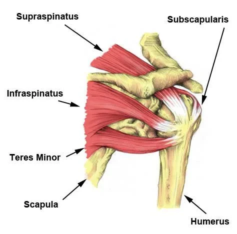
The rotator cuff is a group of 4 muscles that arise from the scapula and insert onto our humeral head, forming a ‘cuff’ around our Glenohumeral joint (shoulder) 


What is the function/role of the Rotator cuff? 

The role of the rotator cuff is to dynamically stabilise the humeral head (Ball) into the glenoid fossa (Socket) whilst the shoulder is moving. Different amounts of rotator cuff muscle recruitment depending on the shoulder movement itself but simply it acts as a ‘stabiliser’ for the shoulder throughout. 

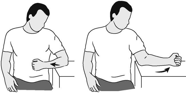
The other, more obvious role, is to rotate the shoulder into internal and external rotation

 Internal Rotation                   External Rotation 


Common Signs and Symptoms of Rotator Cuff Related Shoulder Pain

  1. Pain

Rotator cuff related shoulder pain is generally at the front or on the outside of the shoulder with some cases of referred pain that runs down the outside of our shoulder. Pain can also be apparent on the back of our shoulder blade (scapula)  

  1. Reduced range of motion 

Symptoms can often arise with lifting your arm out to the side (Abduction of the shoulder) or when going into external or internal rotation of the shoulder.Your range of motion is most commonly reduced due to pain 


  1. Weakness and Function impairments 

Tasks that may seem simple can become difficult and painful such as lifting, pulling, overhead movements or reaching behind your back. This can be as a result of pain and weakness from an underlying pathology 

How is Rotator Cuff Related Shoulder Pain Treated? 

Manual Therapy: For those patients with acute or chronic rotator cuff related shoulder pain, a range of manual therapy techniques may be used to help decrease your level of pain, improve your range of motion and improves function. This can include massage, trigger pointing, dry-needling and many more techniques. This is a very important variable of how we treat as physiotherapists but it has an even greater effect when paired with strength and conditioning for the shoulder  


Strength: If one of the contributing factors to RCRSP is muscle weakness or poor function then one of the easiest ways to treat RCRSP is STRENGTHING. 

There is strong, recent evidence to support that strengthening the shoulder has a positive effect on reducing pain and improving function (Dubé, et al - 2020). 

Now to say ‘your shoulder is weak’ is a very global statement that isn’t very specific for our patients. As physiotherapists, our role is to assess and investigate where you have limitations that may be contributing to your RCRSP. From this, specific shoulder exercises will be prescribed for the appropriate loading to the areas of interest within your shoulder. 

Speak to your UPG therapist for a specific shoulder treatment plan

Evidence 

Lewis J. Rotator cuff related shoulder pain: Assessment, management and uncertainties. Man Ther. 2016 Jun;23:57-68. doi: 10.1016/j.math.2016.03.009. Epub 2016 Mar 26. PMID: 27083390.

Dubé M, Desmeules F, Lewis J, et alRotator cuff-related shoulder pain: does the type of exercise influence the outcomes? Protocol of a randomised controlled trialBMJ Open 2020;10:e039976. doi: 10.1136/bmjopen-2020-039976

Teruhisa, M. Chapter: Rotator Cuff Pathology in Textbook of Shoulder Surgery. Jan 2019. doi: 10.1007/978-3-319-70099-1_8.

Written By Dane Monaghan Senior Physiotherapist UPG Studio

Forearm pain associated with prolonged desk work


Forearm pain in desk workers is becoming more prevalent due to the increased time spent working on either the laptop or computer. The two most common causes of forearm pain include overusing the muscles and a poor ergonomic setup of the work environment.

Forearm pain and tightness can lead to referred symptoms down into the wrist and hand as many of the muscles that control your fingers originate in the forearm.  

How do you treat forearm pain?

There are a few treatment options to treat forearm pain and tightness.

  1. Rest the forearm and avoid any aggravating activities

  2. Use an ice pack over the forearm if there is inflammation and swelling observed

  3. Use heat over the forearm muscles if they are feeling tight

  4. Wear a compression garment to provide temporary relief and support

  5. Consult with a physiotherapist for an individualised advice and treatment plan

How do you prevent forearm pain from reoccurring? 

  1. Have a good ergonomic desk set up

  • Is your desk chair at the right height?

    •    If the chair is too high or too low, it will cause pain in the forearm overtime

  • The elbow needs to be at a comfortable 90 deg angle, whether you are standing or sitting. The forearm should be supported

  • Using ergonomically designed keyboards or mouse

2. Minimise sedentary behaviour

  • Regular breaks should be taken throughout the day, especially if your job requires you to be at your desk for the whole duration. Ideally changing positions every hour will reduce the chances of muscle fatigue.

3. Stretch the forearm muscles regularly

  • These stretches can help reduce the stiffness in the forearm, wrist and hands: 

    • Forearm extensors

    •  Have your arm out straight, fingers pointing towards the floor. Add a little pressure to your wrist with your other hand. Hold for 15 seconds. Repeat on the other side.

  • For a deeper stretch, go down onto your hands and knees. Place the back of your hands on the floor, fingers pointing inwards. Hold for 15 seconds

Forearm Flexors 

  • Have your arm out straight, fingers pointing towards the ceiling. Add a little pressure to your wrist with your other hand. Hold for 15 seconds. Repeat on the other side.

For a deeper stretch, go down onto your hands and knees. Place the palms of your hands on the floor, fingers pointing towards your knee. Hold for 15 seconds

Wrist rolls - Roll your wrist clockwise and anti clockwise.

4. Strengthen the forearm muscles

  • Resistance exercises are important to build the strength in the forearm muscles and prevent the pain from occurring. Choose an appropriate weight for the following exercises. Usually a 0.5kg dumbbell is a good starting weight. 

  • Wrist flexion - Resting your forearm on the table with your hand hanging off the edge, palms facing upwards, bend the weight inwards x 10

  • Wrist extension - Resting your forearm on the table with your hand hanging off the edge, palms facing downwards, bend the weight inwards x 10

  • Wrist pronation/supination - Turning your wrist over x 10

Our Physiotherapists at UPG studio have experience in the assessment, treatment and management of forearm pain. We ensure a comprehensive assessment is completed not just for the symptoms presented, but also to address the underlying causes and contributing factors. Our detailed rehabilitation plans are tailored to each individual to ensure the best possible outcome.


To stretch or not to stretch?

Stretching is often a neglected component of fitness but its role in maintaining optimal health is more important than you think. You may not know it, but our muscles are always working whether, for example, passively to keep you upright or actively when we exercise. This means that from postural habits to exercise or as a normal part of ageing and disuse, our muscles will shorten, build up tension and become tight over time consequently reducing the range and weakening the muscle. Unfortunately, this increases your risk of injury such as strains, joint pain and muscle damage. Not to worry because this is where stretching comes into play!

Simply put, stretching lengthens your muscles. This increase in flexibility helps to maintain and improve the range of motion in your joints which ultimately reduces your risk of injury. It can also optimise athletic performance, reduce pain and alleviate post exercise muscles soreness. Stretching’s low impact nature makes it is suitable for everyone. It feels good and it is relaxing. Stretching can be the gateway into improving your wellbeing and being more mindful. And this is especially important now given all the built-up pandemic stress and stress from balancing work-life commitments as life returns to normal.

Stretching is a little bit more complicated than trying to touch your toes. There are 3 main types of stretching. These are static, dynamic and pre-contraction stretching, each with a different technique and are used for different purposes. It is important to know when a particular type of stretching exercise is most appropriate. Recruiting a physiotherapist can help set you up with a tailored program to meet your needs and maximise your goals.

-        Static stretching involves holding a stretched position for a long period of time greater than 10 seconds. This form of stretching increases your muscle length, helps reduce the risk of muscle injuries and aids recovery. Static stretching should be avoided when warming up as it can hinder your performance.

-        Dynamic stretching requires you to move through a range of motion in a repetitive manner. It primes the body for exercise to help protect you from injury and can decrease the soreness felt post workout.

-        PNF (Proprioceptive Neuromuscular Facilitation) stretching is the most common type of pre-contraction stretching. PNF stretching’s 3 different techniques facilitate a passive stretch by either activating the agonist or antagonist muscle. This method of stretching will have a more immediate result regarding changes in flexibility and joint range compared to the other forms of stretching and can induce increases in strength, power and consequently performance.

We do not need to achieve gymnast level flexibility to keep our muscles strong and healthy. Rather, we need to aim for balance between the left and right and the front and back of our bodies!

Here are my top 3 stretches

-        Glute (figure 4)

1)     Lie on your back with your knees bent and feet on the floor (crook lying)

2)     Rest your right ankle over your left knee, making a figure 4

3)     Hug your left knee and bring it towards your chest

-        Lower back (windshield wipers)

1)     Lie on your back with your knees bent and feet on the floor, arms out to the side like a T

2)     Keeping your feet on the ground, slowly lower your bent knees to the left and turn your gaze to the right.

-        Neck (upper trap)

1)     Bring your ear to your shoulder whilst keeping your shoulders relaxed, down and back

According to guidelines stretching 2 -3 times a week confers the best results! Happy stretching!

Page P. (2012). Current concepts in muscle stretching for exercise and rehabilitation. International journal of sports physical therapy, 7(1), 109–119.

Stretching. (2022, March 31). Physiopedia, . Retrieved 07:19, April 3, 2022 from https://www.physio-pedia.com/index.php?title=Stretching&oldid=299382.

Hindle, K. B., Whitcomb, T. J., Briggs, W. O., & Hong, J. (2012). Proprioceptive Neuromuscular Facilitation (PNF): Its Mechanisms and Effects on Range of Motion and Muscular Function. Journal of human kinetics, 31, 105–113.

Chaabene, H., Behm, D. G., Negra, Y., & Granacher, U. (2019). Acute Effects of Static Stretching on Muscle Strength and Power: An Attempt to Clarify Previous Caveats. Frontiers in physiology, 10, 1468.

O'Sullivan, K., Murray, E. & Sainsbury, D. The effect of warm-up, static stretching and dynamic stretching on hamstring flexibility in previously injured subjects. BMC Musculoskelet Disord 10, 37 (2009).

Written by Jenny Wu : Physiotherapist - UPG Studio

Iliotibial Band Syndrome

What is the iliotibial band?

The iliotibial band is a connective tissue that is located along the outer thigh and knee. It works to provide strength and stability to the front and side of the knee. It also helps to stabilise the hip during walking, running and hopping. 

The band is made up of a strong fibrous material which can work like a spring in absorbing and releasing energy. 

What is iliotibial band syndrome (ITBS)?

Iliotibial band syndrome is a common knee injury that typically presents as pain or tenderness on the outside of the knee. It is often due to overuse or a rapid change in training volume but may also be due to associated muscle weakness of the surrounding structures. This leads to compression and inflammation of the iliotibial band. It occurs most often in cyclists, long distance runners and athletes who frequently squat.

Iliotibial band syndrome is caused by a combination of factors, including:

  • Poor training habits or technique

  • Sudden increases in activity levels

  • Reduced muscle flexibility

  • Muscle weakness 

  • Mechanical imbalances in the body - often in the lower back, pelvis, hips and knees 

What are the signs and symptoms of ITBS?

  • Sharp pain on the outer side of the knee

    • Particularly during running when the heel strikes the ground

  • Sensation of stinging or prickling 

  • Snapping or popping sensation at the knee

  • Pain aggravated by movements which load the knee e.g. squats, running, stairs

  • Pain radiating upwards along the length of the ITB to the outer thigh and hip 

How is it diagnosed in physiotherapy?

Physiotherapists will first ask questions to understand the potential causes of your symptoms. This can include questions about your training history, symptom presentation as well as lifestyle and activity. 

There is also a physical exam where physiotherapists will perform key assessments and look at your range of movement, movement control, strength, posture and gait.

Recovery 

Most people recover from iliotibial band syndrome. However, it may take weeks or months to return to full activity without pain. 

How can physiotherapists at UPG help treat your iliotibial band syndrome? 

We can help by:

  • Reducing acute pain and providing advice on what to do during the early stages

  • Providing education about training volume, running form and technique

  • Addressing the causes contributing to iliotibial band syndrome e.g. weakness, pain, reduced flexibility, posture

  • Prescribing appropriate exercises as well as strength and conditioning work

  • Guiding load management and return to running/activity safely

  • Progressing your rehab to help you achieve your goals 

Book in with one of our friendly UPG physiotherapists for a consultation. Together, we can help you get back to doing the activities and hobbies that you love to do!

References:

Charles, D., & Rodgers, C. (2020). A Literature Review and Clinical Commentary on the Development of Iliotibial Band Syndrome in Runners. International Journal of Sports Physical Therapy 15(3), 460-470.

Jerold M. Stirling et al., (2015). Iliotibial Band Syndrome Treatment & Management. https://emedicine.medscape.com/article/91129 


​​Plantar Fasciitis

​​Plantar Fasciitis

What is it?

Plantar fasciitis is inflammation of the plantar fascia, a piece of connective tissue that runs along the bottom of the foot. Plantar fasciitis is the most common cause of heel pain (Bahr, 2012). Usually It is developed after overuse, over stretching or from a medical condition.


What are the common symptoms?  

-   Pain under the heel

-   Usually bothersome in the morning when getting up from bed, after prolonged sitting/standing or after intense physical activity (Sports Medicine Australia,2022) .


What can increase my chances of developing plantar fasciitis?

-   Physical activity with high stress on the heel bone, e.g. running.

-   Being overweight

-   Pregnancy

-   Feet deformities

-   Wearing ill fitting shoes

-   Tight calf muscles

-   Above middle age

-   Being on your feet for prolonged periods (Sports Medicine Australia, 2022)


How will this be diagnosed?  

-   Physiotherapists are usually able to make a diagnosis after asking you some questions about your symptoms, type of work you do and your lifestyle.

-   There will also be a physical exam where the therapist will have a look at your feet checking for swelling, redness, any points of tenderness, stiffness, tightness or weakness.


What are the different types of treatment options?

-   There are a few interventions a physiotherapists can guide you through including:

  • Recommendation for pain-relief medications

  • Modifications to your training/weight bearing activity

  • Modifications to footwear or insoles

  • Stretching exercises specific for plantar fasciitis

  • Friction massage

  • Taping

  • And therapeutic cold ice recommendations  


Will my pain go away?

  • Usually the pain can improve over time with conservative treatments

  • Most people become asymptomatic within 4-6 weeks (Bahr, 2012)


Why come to UPG?

  • If you are suffering from heel pain similar to this come to see one of our physiotherapists to help guide you through your rehabilitation and treatment. The physiotherapists at the clinic will be able to assist with:

  • Reducing the initial pain and inflammation

  • Improve your flexibility

  • Strengthen the plantar fascia

  • Return to play

  • Prevent this from happening again

    Written by Phoebe Cunningham : Physiotherapist - UPG Studio

References:

Bahr, R. (2012). The IOC manual of sports injuries: An illustrated guide to the management of injuries in physical activity. John Wiley & Sons.

Health Direct. (2021, June 20). Plantar fasciitis. Trusted Health Advice | healthdirect. https://www.healthdirect.gov.au/plantar-fasciitis

Sports Medicine Australia. (2022). Plantar Fasciitis. https://sma.org.au/resources-advice/injury-fact-sheets/plantar-fasciitis/


Acute Wry Neck

What is Acute Wry Neck?

Acute wry neck is a very common condition that often presents with a sudden onset of pain and stiffness in the muscles around your neck commonly accompanied by tightness or spasm in the surrounding neck muscles. This muscle spasm can be a protective response that can cause pain and the inability to move the neck (Cohen, 2017). Around two-thirds of the population will experience neck pain at a given point in their life (Vos, 2007). Anyone can get a wry neck, but it typically occurs in young people between 12 and 49 years of age (Counsell, 2016).

Causes of Acute Wry Neck

The cause may not always be known but there is a strong connection to sleeping in an abnormal position without proper head and neck support, inappropriate seating, poor posture while at a desk, or carrying heavy unbalanced loads. The onset of the pain and stiffness is usually sudden either caused by the locking of a facet joint or irritation to a disc in the neck with the most common cause being a locked facet joint (Groeneweg, 2017). The significant pain from a wry neck is explained by the high density of nerve fibers in the tissues surrounding these joints.

What are the Risk Factors? (Brukner & Khan, 2017)

  • People aged 12-49 years old

  • Occupations such as office workers

  • Sleeping disorders

  • Depression or anxiety

  • A previous history of neck pain

  • Women are more prone to neck pain than men

  • Sedentary lifestyles

  • Smoking

  • Being overweight or obese

Signs and Symptoms of Acute Wry Neck (Brukner & Khan, 2017)

  • Pain: The onset of pain is sudden and commonly located in the middle or side of the neck that is affected most. The pain is localised to the neck area and does not extend past the shoulder joint.

  • Location: Usually symptoms are felt on one side of the neck, as this is a protective reaction of the body to safeguard the neck from further injury or damage.

  • Loss of Range of Movement: The neck is often fixed in an abnormal position (most frequently in a flexed forward and rotated position). The side that the patient’s head is rotated towards will often be away from the side of pain due to this protective reaction.

  • Muscle Spasms: The surrounding neck muscles often have tightened or spasmed in response to the facet joint irritation. This in turn limits the neck range of motion.

  • Referring Pain: Generally, there is no referring pain however Discogenic wry necks often present in the same way but are more likely to travel down the arm and may experience pins and needles in their hand as well.

Prognosis 

The prognosis for a wry neck is generally very good and with the correct diagnosis and timely treatment, people will often feel relief within 2-3 sessions and return to pre-injury function within a week (Vos, 2007). Discogenic wry neck also has a good prognosis and will often feel a lot better within the first few sessions however, can take 2-6 weeks to return to pre-injury function depending on the severity (Brukner & Khan, 2017; Vos, 2007).

Rehabilitation and Prevention 

Early management for both facet and discogenic wry neck with your physiotherapist will often be gentle and involve facet joint mobilisation and soft tissue release of the surrounding neck muscles. 

Common early management recommendations include the use of heat packs for pain relief as well as re-introduction of gentle neck movements and avoidance of heavy lifting and rapid head movement. Once the pain has settled significantly and movement is mostly restored, prevention involves exercises targeted at strength and endurance for muscles surrounding the neck (Brukner & Khan, 2017; Vos, 2007).

It’s worth checking your pillow to make sure that your head and neck are well supported while sleeping and avoid sleeping on your belly whilst pain is present. Avoid using a rigid neck brace, you want to keep your head and neck mobile to reduce any further associated stiffness.

Common treatment methods which your physiotherapist may recommend to settle acute wry neck include:

  • Massage

  • Dry needling

  • Gentle passive mobilisations

  • Heat therapy

  • Stretches/ light movement

  • Non-steroidal anti-inflammatory medication

Wry neck which is not treated can result in stiffened neck segments which can lead to an individual being predisposed to recurring episodes of this condition. 

Our Physiotherapists at UPG Studio have experience in the assessment, treatment, and management of many conditions including Wry Neck. We ensure a comprehensive assessment is completed not just for the symptoms presented, but also addressing the underlying causes and contributing factors. Our detailed rehabilitation plans and Physfit programs are tailored to each individual to ensure the best possible outcome. Always consult your physiotherapist or medical professional for an assessment of your condition before taking on any new exercises.

References
- Brukner, P., & Khan, K. (2012). Clinical Sports Medicine, 4th Edition
- Cohen, S. P. (2015, February). Epidemiology, diagnosis, and treatment of neck pain. In Mayo Clinic Proceedings (Vol. 90, No. 2, pp. 284-299). Elsevier.
- Cohen, S. P., & Hooten, W. M. (2017). Advances in the diagnosis and management of neck pain. Bmj, 358.
- Counsell, C., Sinclair, H., Fowlie, J., Tyrrell, E., Derry, N., Meager, P., … Grosset, D. (2016). A randomized trial of specialized versus standard neck physiotherapy in cervical dystonia. Parkinsonism & related disorders., 23, 72–9.
- Groeneweg, R., van Assen, L., Kropman, H., Leopold, H., Mulder, J., Smits-Engelsman, B. C., ... & van Tulder, M. W. (2017). Manual therapy compared with physical therapy in patients with non-specific neck pain: a randomized controlled trial. Chiropractic & manual therapies, 25(1), 1-12.
- Vos, C., Verhagen, A., Passchier, J., & Koes, B. (2007). Management of acute neck pain in general practice: a prospective study. British journal of general practice, 57(534), 23-28.


Muscle strains

What are muscle strains?

Muscles are strained (or torn) when some or all of the muscle fibres fail to cope with the demands placed upon them. Muscle strains are most commonly associated with sporting injuries – typically when either accelerating or decelerating suddenly. Activities that require higher speeds or of a stop-start nature tend to produce a higher number of muscle strains. Muscle strains are also very common at the workplace, during activities of daily living and with incidents such as falls/slips.

All strains involve the tearing of a number of fibres of the muscle, evoking both pain and swelling. Tears are graded from 1 through to 3 in terms of their severity. The most common sites of muscle strains tend to be muscles that cross 2 joints, e.g. the hamstrings, calves, quadriceps, adductor (groin) muscles but can occur in almost any muscle in the body.

What can I do if I strain a muscle?

An accurate diagnosis, early management and specific rehabilitation of muscle strains are crucial in getting you back to normal activity and preventing reinjury. A lot of the time muscle strains are treated poorly with many people believing that once the pain has subsided the muscle has healed. This however is not the case. Pain will often resolve after the first 1-2 weeks (depending on severity) following injury, but it can take weeks more to regain muscle function back to normal. If poorly rehabilitated, the muscle fibres will fail to heal adequately and as a result normal muscle function is unlikely to return placing it at risk of re-straining.

Early and appropriate management is the key to the best outcomes. How well you manage your injury in the first few days can make a big difference in recovery time and prevent reinjury. The initial management for a muscle strain as with all soft tissue injuries should follow the R.I.C.E.R protocol.

Rest - Protect damaged tissue from more injury by stopping activity. Use crutches (or sling for upper limb strains) as needed. Immobilise to reduce pain and bleeding, and prevent disruption of the healing process. Rest for 3-5 days depending on severity of injury.

Ice - Apply ice to decrease pain and muscle spasm. Ice with the injured muscle on slight stretch (pain free) to help prevent ice scarring. Ice for 20 minutes and repeat every 2 hours on the first day and then every 4 hours on the second day and for at least 3 days after injury.

Compression - Apply compression to the area to reduce swelling, ensuring that it isn’t too tight that it impedes blood flow to the end of the limb or cause pins and needles or numbness.

Elevation - Elevate the injured limb above the level of the heart as much as possible in the first 72 hours to assist with the swelling.

Referral - Seek advice from an expert therapist. Your Physiotherapist is experienced in the assessment, treatment and management of muscle strains. Early and appropriate treatment and advice is recommended for optimum recovery and prevention of repeat injuries.

What not to do!

No heat, alcohol, running or massage in the acute healing phase. All these factors will increase swelling and bleeding in the injured area and delay the healing process.

How do I prevent muscle strains?

Most muscle tears are preventable. Prevention requires good management of predisposing factors such as:

  • Inadequate warm up

  • Excessive muscle tightness and insufficient joint range of movement

  • Muscle fatigue, over use, inadequate recovery

  • Muscle imbalances

  • Previous injury

  • Faulty technique/biomechanics

  • Spinal dysfunction (including poor core stability)

Our Physiotherapists at UPG studio have experience in the assessment, treatment and management of muscle strains. We ensure a comprehensive assessment is completed not just for the symptoms presented, but also addressing the underlying causes and contributing factors. Our detailed rehabilitation plans and physfit programs are tailored to each individual to ensure the best possible outcome

 Written by Erin Gleadell: Pilates Lead/Physiotherapist UPG Studio

 References:

Heiderscheit, B., Sherry, M., Silder, A., Chumanov, E. and Thelen, D., 2010. Hamstring Strain Injuries: Recommendations for Diagnosis, Rehabilitation, and Injury Prevention. Journal of Orthopaedic & Sports Physical Therapy, 40(2), pp.67-81.

Fernandes, T., Pedrinelli, A. and Hernandez, A., 2011. MUSCLE INJURY – PHYSIOPATHOLOGY, DIAGNOSIS, TREATMENT AND CLINICAL PRESENTATION. Revista Brasileira de Ortopedia (English Edition), 46(3), pp.247-255.


Running and Patellofemoral Pain Syndrome

The Knee to Running!

Running has one of the largest participation rates, a survey found that about 1,224,100 Australians aged 15 years and older participated in running. As the Melbourne Run is coming up, I believe many people are having the urge to join the race in light of it being one the largest sporting events we can participate in after the easing of restrictions.

Some of you might have been training all throughout lockdown but I would anticipate that many would have just started a few weeks ago going out for a 5km run after 6 months of limited physical activity and ending up having sore knees, feet and ankles. Does this sound like you? If so, keep reading! 

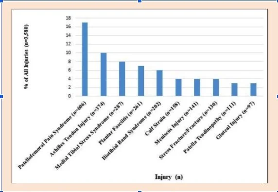
According to a recent study (Francis et al, 2018), they have concluded that the top 3 injuries are:

  1. Patellofemoral Pain Syndrome (PPS)

  2. Achilles Tendon Injury

  3. Medial Tibial Stress Syndrome

In this blog I will mainly focus on patellofemoral pain syndrome aka anterior knee pain syndrome.

What is Patellofemoral Pain Syndrome (PPS)?

Amongst the patients from a sports clinic,  70% of diagnosed cases of PPS are aged 16–25 years (Devereaux and Lachmann, 1984).  This condition also tends to affect women more than men by 25% (Boling et al, 2010)

Signs & Symptoms:

  • Knee pain with squatting, running and walking down stairs (Cook et al, 2010)

  • Described as pain behind the knee 

  • Pain worse after activity (Petersen et al, 2014)

  • Anterior knee pain with prolonged sitting “theatre sign” (Larson et al, 1978)

Why does my knee hurt?

When we are running, each step we take roughly pushes back 2.5 times body weight into our foot (Hamil et al, 1983). Being the main shock absorber when running, our knees take up most of the forces, 4 times body weight to be exact (Willy et al, 2016). Translating this to a full marathon, let’s assume you are able to complete 42kms in approximately 4 hours and 20 minutes with a step rate of 160 steps per minute, your knees taking a total of 320 body weights per minute: 

320 x 260mins = 83,200 times the body weight !!! (Neumann, 2002)

With such a large amount of load going through your knees, if your body does not have the capacity to recover between training sessions the end result is overtraining and overuse of the knees leading to PPS.

So what can you do about it?

There are 2 main thoughts behind managing PPS. 

First, is reducing the load:

  • Adjusting training to maintain pain levels at a maximum of 2/10 on a numeric pain rating scale and increasing based on symptoms

  • Modifying running pattern by increasing step rate and changing foot-strike pattern (Esculier et al, 2018)

Second, is to improve our capacity of the knees to take more load:

  • Knee taping to promote better tracking of the knee cap 

  • Combination strengthening exercises targeting the knees, hip and trunk muscles (Colins et al, 2018)

With the assistance of our enthusiastic physiotherapist team at UPG, together we can take on the recovery of your sporting overuse of acute injuries so that you can go back to enjoying your passion care-free and pain free. Book in for your initial assessment before your pain and symptoms worsen.

 Written by Jason Lui: Physiotherapist UPG Studio

References:

  1. Boling M, Padua D, Marshall S, Guskiewicz K, Pyne S, Beutler A. Gender differences in the incidence and prevalence of patellofemoral pain syndrome. Scand J Med Sci Sports. 2010;20:725–30

  2. Collins NJ, Barton CJ, van Middelkoop M, et al. 2018 Consensus statement on exercise therapy and physical interventions (orthoses, taping and manual therapy) to treat patellofemoral pain: recommendations from the 5th International Patellofemoral Pain Research Retreat, Gold Coast, Australia, 2017. Br J Sports Med. 2018;52(18):1170–1178. doi: 10.1136/bjsports-2018-099397

  3. Cook C, Hegedus E, Hawkins R, Scovell F, Wyland D. Diagnostic accuracy and association to disability of clinical test findings associated with patellofemoral pain syndrome. Physiother Can. 2010;62(1):17-24.

  4. Devereaux MD, Lachmann SM.  Patello-femoral arthralgia in athletes attending a sports injury clinic. Br J Sports Med. 1984;18:18–21

  5. Esculier JF, Bouyer LJ, Dubois B, et al. Is combining gait retraining or an exercise program with education better than education alone in treating runners with patellofemoral pain? A randomised clinical trial. Br J Sports Med. 2018;52(10):659–666. doi: 10.1136/bjsports2016-096988

  6. Francis P, What,an C, Sheerin K, Hume P, Johnson MK. The proportion of lower limb running injuries by gender, anatomical location and specific pathology: a systematic review. Journal of Sports Science and Medicine. 2018;18: 21-31

Don’t underestimate ankle sprains!


Over my 20 year career as a physiotherapist, I’ve seen a fair share of ankle sprains in private practice, community care and in residential aged care. 

Ankle sprains are extremely common and are almost seen as “routine” in sport, especially those involving changes of direction. Ankle sprains are also common when elderly people overstep and fall and can lead to longer recovery times and deterioration in their standing balance. 

Unfortunately, these injuries are often not rehabilitated completely and can lead to all sorts of continual problems. Anatomically as a refresher, most commonly injured are the ligaments on the outside of the ankle, namely the specific band called the anterior talo-fibular ligament, or ATFL for short. This band of fibrous tissue limits plantarflexion (pointing the foot down) and inversion (turning the foot inwards). Luckily, should it be damaged through an ankle sprain, it has a good capacity to heal and for scar tissue and there are other surrounding ligaments which still provide good stability.

Here’s a trap for young players: beware of just resting and waiting for a simple ankle sprain to resolve. A number of studies have shown that high level balance and proprioception (ability to sense and react to various positions in standing) DO NOT come back on their own (Hiller et al., 2011). 

This leaves the athlete and particularly the elderly patient with a potentially unstable platform and the consequences can vary, including:

  • Recurrent sprains, leading to chronic instability and early degenerative changes

  • Altered movement patterns

  • Compensation further up the “chain” ie: knee or hip problems which also increases falls in the elderly

  • Increased risk of acute knee injury, such as an ACL rupture in the athlete when returning back to competitive games

At UPG Studio, our physiotherapists can provide a well-designed rehabilitation and reablement programs (Postle, Pak and Smith, 2012) which is critical and help to avoid recurrence and reduce the risk of the above.  We have specific reablement programs designed by physiotherapists at UPG Studio that focuses on enhancing an individual’s rehabilitative potential through the prescription of active and short-term restorative programs which have been shown to provide many benefits to the older people living at home as well as athletes.

James at work in the studio, with an elite athlete.

These restorative programs include restoring full movement and strength which is a given. But close attention should be given to proprioceptive and neuromuscular retraining. My best three high level balance exercises are included below. (Please note, these should only be done when pain and swelling have subsided and please consult a physiotherapist for further guidance if required)

Bosu ball drills

  • Standing on the injured side and balancing on the flat side

  • Hopping and rotating on the ball side 

Hopping and landing challenges

  • Create challenges such as jumping over a stick and landing on a mark

  • Jumping and landing between two objects 

Shuttle runs

  • Make sure you turn through both directions, with increasing speed

Our Physiotherapists at UPG studio have experience in the assessment, treatment and management of ankle sprains. We ensure a comprehensive assessment is completed not just for the symptoms presented, but also addressing the underlying causes and contributing factors. Our detailed rehabilitation plans and physfit programs are tailored to each individual to ensure the best possible outcome.

Written by James Nguyen Founder, Managing Director & Head Physiotherapist- UPG Studio

References:

Characteristics of people with recurrent ankle sprains: a systematic review with meta-analysis (Claire E Hiller, Elizabeth J Nightingale1, Chung-Wei Christine Lin, Garrett F Coughlan, Brian Caulfield, Eamonn Delahunt) - Br J Sports Med doi:10.1136/bjsm.2010.077404 

Effectiveness of proprioceptive exercises for ankle ligament injury in adults: A systematic literature and meta-analysis - K. Postle, D. Pak, T.O. Smith, - Manual Therapy

Volume 17, Issue 4, August 2012, Pages 285–291 

About James Nguyen

James is the Managing Director and Head Physiotherapist at United Physiotherapy Group. 

With over 20 years experience in private practice & sporting settings, he is dedicated to the holistic management of complex chronic spinal, pelvic and lower limb issues.

Chronic Neck Pain

Chronic neck pain

The chance that you will have neck pain at some point over your lifetime is nearly 50%.

This tends to be most prevalent in middle aged people.

On most occasions, an acute episode of neck pain with treatment from a physio will resolve in 2-8 weeks (very case dependent) however, CHRONIC neck pain can persist in 10-30% of people.1

Chronic neck pain is pain that lasts for 12 weeks or more and is often associated with restricted movement, associated pain, muscle or joint tightness and often sore to touch.

Causes / Risk Factors

  • Sustained postures or movements that strain the neck

  • Very common in occupations which require a lot of work with their arms above shoulder height

  • Roles with exposure to conflict or high stressors

  • Office/desk workers with poor workspace ergonomics i.e. looking down for long periods of time 2 

How to fix it and avoid neck pain from ccurring

Manual therapy (Joint-mobilisations) + an exercise program produces greater long-term improvement in neck pain compared to other forms of treatment. This includes conditions like cervicogenic headaches (neck-related headaches), chronic neck pain/stiffness and neck pain with associated radicular or referred pain i.e. neck pain + pain down your arm or into your shoulder blade for example4

Strength and mobility exercises to target and offload specific structures around the neck and shoulder are important. We train many muscles of the body but neck or upper back muscles are commonly left untrained. Weakness, poor technique or overuse to muscles of our neck can lead to the joints of our neck being exposed to higher loads than they can manage which often leads to pain.

Workplace ergonomic assessments 

Due to the recent increase of working from home, our desk or workstation setup may not be as ergonomically friendly to our neck and upper back region. Quite often forward neck postures are being adopted which are placing higher stressors onto the surrounding muscles, ligaments and joints of our neck causing pain 2.

Managing stressful situations 

When the body is stressed, muscles tense up, it’s the body's own defence mechanism against stress to guard you from injury or pain. Once stress passes, the muscle tension releases. When this becomes problematic is when we have chronic stresses being experienced meaning that muscles are in almost a constant state of protection. When this occurs, this can trigger other reactions in the body secondary to the initial reaction to stress.

Cervicogenic headaches / Neck related headache

A headache can come from a number of different causes and can often be missed. If the following is sounding familiar to you or someone you know then you/they may be suffering from a neck related headache or more commonly known as a Cervicogenic Headache.

A cervicogenic headache is the most common cause of chronic headaches.

Symptoms

  • One sided headache

  • Reduced neck range of motion

  • Tenderness on upper neck (C1-C3)

  • Tightness in surrounding muscles

INTERESTING FACT

Our C1-C3 nerves relay pain (nociception) signals to a specific area of the head and neck called the ‘trigeminal nucleus’. This connection can have the output of referred pain to some of our facial nerves which helps give us the feeling of a ‘headache’.

**Please note this is a very brief explanation of a very complex system**  

CAUSES

Referred pain from an irritation of structures innervated by spinal nerves C1-C3.

So what does this mean?

Structures around the upper part of your neck such as the joints, disc, ligaments and muscles can be dysfunctional in different ways which then activates the specific nerves associated.

The most common example is our facet joints of C1-C3 (top of your neck) being stiff and sore. Like any spinal injury this can be acute or chronic in nature.4

Our spinal nerves C1-C3 messages share the same region as our facial nerves so when we have an irritation to these structures the output can be referred pain to some of our facial nerves resulting in a headache! Weird isn’t it!

TREATMENT

If the cause is likely from the facet joints being stiff and sore within your neck, then the obvious treatment is through manual therapy to those joints affected.

Manual therapy can reduce pain, reduce stiffness with a joint and improve function. Once we begin to help increase our joint mobility around the affected joint then there is less stress on our C1-C3 nerves which should result in REDUCED HEADACHES! 5

Following Manual therapy, specific exercises are likely to be prescribed to help prevent this reoccurring again. 

HOW WE CAN HELP AT UPG 

Firstly, our UPG physios can identify and treat chronic neck pain and the potential associated headaches. Treatment varies from person to person but our very skilled physios excel with their manual therapy skills to make you feel better than ever. Once symptoms have been relieved, then specific exercises will be prescribed to ensure that this type of pain can stay away.

At UPG we are happy to help, don’t suffer from chronic neck pain and associated headaches any longer. Book in for your initial assessment before your pain and symptoms worsen.

Written by Dane Monaghan: Physiotherapist UPG Studio

References 

1. Hogg-Johnson S, Van der Velde S, Carroll L, Holm L, Cassidy D, Guzman J, Côté P, Haldeman S,
Ammendolia C, Carragee,14,15 Eric Hurwitz E, Nordin, M, Peloso P. The Burden and Determinants of Neck Pain in the General Population: Results of the Bone and Joint Decade 2000–2010 Task Force on Neck Pain and Its Associated Disorders. Eur Spine J. 2008 April; 17(Suppl 1): 39–51.

2. Ostergren PO, Hanson BS, Balogh I, Ektor-Andersen J, Isacsson A, Orbaek P, Winkel J, Isacsson SO; Incidence of shoulder and neck pain in a working population: effect modification between mechanical and psychosocial exposures at work? Results from a one year follow up of the Malmö shoulder and neck study cohort. Malmö Shoulder Neck Study Group. J Epidemiol Community Health. 2005 Sep; 59(9):721-8. 


3. Al Khalili Y, Jain S, Murphy PB. 2019 Headache, Cervicogenic

4. Becker WJ. Cervicogenic Headache: Evidence that the neck is a pain generator. Headache. 2010;4 699-705

5. Fritz JM, Brennan GP. Preliminary Examination of a Proposed Treatment-Based Classification System for Patients Receiving Physical Therapy Interventions for Neck Pain. Physical Therapy. 2007;87:513-524.

Physical Activity and Exercise Recommendations for Adults

As we continue to face the ongoing impacts of COVID-19, our normal routines have been thrown into chaos and it is easy to forget our daily active routines that we once had. This blog post is a little reminder of what we need to do to improve our physical health and mental wellbeing.

  • Studies show that maintaining a regular active lifestyle can:

  • Reduce the risk of type 2 diabetes and cardiovascular disease

  • Improve and maintain blood pressure, cholesterol and blood sugar levels

  • Maintain strong muscles and bones

  • Maintain a healthy weight

  • Improve mental health by having an antidepressant effect in people with mild to moderate depression.

  • Improve sleep quality

(Department of Health, 2021; World Health Organization, 2020)

How much and what should we be doing?

The World Health Organization recommendations for physical activity in adults each week include at least:

  • 150 minutes (2.5 hours) of moderate intensity physical activity OR

  • 75 minutes of vigorous intensity physical activity In conjunction with 2 sessions of muscle-strengthening training sessions

Moderate intensity physical activity examples include brisk walks, golf, swimming.

Vigorous intensity physical activity examples include running, soccer, netball, fast cycling and most competitive sports.

Muscle-strengthening exercise examples include weight-lifting, reformer Pilates or any activity which involves lifting or carrying. These resistant training sessions should target all the major muscle groups within the body for maximal benefits.

It is important to remember that any type of exercise is better than none. Making small changes in your daily routine will make a difference in building long lasting habits. For example, instead of driving to the grocery store, choose to walk or ride a bike instead.

How can a physiotherapist help me?

Physiotherapists are experts at helping our clients create a tailored exercise plan. We consider our clients’ interests, current ability, goals and any pre-existing injuries they may have. It is important to develop a routine which is safe, appropriate and enjoyable for you, otherwise the likelihood of long-term adherence and maintenance will be low.

At our UPG South Yarra Studio, we offer Physfit reformer Pilates to our clients as a structured strengthening regime tailored to your needs. 

If you have any concerns or would like to improve your current exercise routine, please get in touch with the team at UPG to book an appointment with one of our physiotherapist.

Written by Anna Diep - UPG Physiotherapist and Pilates Instructor

References:

Department of Health. (2021). Physical activity and exercise guidelines for all Australians. https://www.health.gov.au/health-topics/physical-activity-and-exercise/physical-activity-and-exercise-guidelines-for-all-australians

Bull, F. C., et al. (2020). World Health Organization 2020 guidelines on physical activity and sedentary behaviour. British journal of sports medicine, 54(24), 1451-1462, https://bjsm.bmj.com/content/bjsports/54/24/1451.full.pdf

Pregnancy and Pilates

It is recommended that you check with your doctor, midwife or physiotherapist prior to starting an exercise program throughout your pregnancy.

If you do not have any medical obstetric complications, you will be able to continue to exercise throughout your pregnancy. However, your exercise regime should be modified throughout each stage of your pregnancy.

Modifications to your exercise program. 

Throughout your pregnancy, you may need to reduce the intensity of your exercise program. Exercise should be kept to a light to moderate intensity. Physical Activity Australia (2020) recommends that the ‘talk test’ should be used as a guide to measure the intensity of the exercise. This means that you should be able to maintain a conversation comfortably whilst exercising. Australian Physical Activity Guidelines recommends 150 minutes of moderate intensity exercise per week for pregnant women. However, it is important to listen to your body and exercise within your own limits. There are specific recommendations for modifications to make to your exercise regime for each trimester which is outlined further below.  

You should be monitoring your heart rate and exertion throughout your exercise. Physical Activity Australia (2020) recommends that pregnant women should exercise at less than 75% of their maximum heart rate. The Royal Australian and New Zealand College of Obstetricians and Gynaecologists (RANZCOG) (2020) recommends that you should avoid increasing your body temperature. Therefore, exercise should be reduced on hot or humid days and you should maintain adequate hydration and wear loose, breathable clothing.

You should avoid exercising flat on your back after the first trimester. Besides being uncomfortable and causing you dizziness, laying flat on your back places the uterus directly onto the vena cava which is a major vein that returns blood to the heart. This can compromise the return of blood to the heart which can make you feel dizzy and nauseous. Instead, you can prop some pillows under your back or try to practice these exercises on your side.

When you should stop exercising:   

If you experience any of the below signs, immediately stop your workout and seek advice from your obstetrician.

  • Chest pain

  • Unexplained shortness of breath

  • Dizziness, feeling faint or headache

  • Muscle weakness

  • Calf pain, swelling or redness

  • Sudden swelling of ankles, hands or face

  • Vaginal bleeding or amniotic fluid loss

  • Decreased fetal movement

  • Uterine contractions or pain in the lower back, pelvic area or abdomen

(The Royal Australian and New Zealand College of Obstetricians and Gynaecologists, 2020).

Exercising throughout your pregnancy.

Evidence suggests that pregnant women should continue to participate in regular aerobic and strength conditioning (RANZCOG, 2020). Exercising during pregnancy helps to protect your body against strain and injury, boosts circulation of blood and lymphatic fluids, keeps your muscles toned and can have many psychological benefits.

Exercising in the 1st trimester.

The first trimester is where the most foetal development occurs and it is also the time where miscarriage is most likely to occur (Raising Children Network (Australia) Limited, 2020). Exercise during this time should be gentle. Pilates during this stage of pregnancy can assist with providing a strong centre which can act to brace the spine, support the extra weight from the baby and provide extra stability to your joints. Pilates also assists with strengthening of the pelvic floor which will help to support the uterus and reduce stretching of the pelvic floor during and after pregnancy.

Exercising in the 2nd trimester.

As your uterus is now enlarged, it is recommended that in the 2nd and 3rd trimester you should avoid performing exercises flat on your back for long periods of time (RANZCOG, 2020). Instead you can prop your back up with pillows or practice these exercises on your side.

Exercising in the 3rd trimester.

At this stage of your pregnancy you are carrying roughly an extra 12kg of weight. You will most likely feel more tired and breathless. There may be more fluid (oedema) in your hands and feet and due to your increase in uterus size there will be less room within your digestive system, which may cause constipation and heartburn. Gentle Pilates exercises can help with these problems.

If you are wanting to exercise throughout your pregnancy, get in touch with the team at UPG where one of our physiotherapists can perform a specific assessment and create an exercise program suited to your needs. 

Written by Phoebe Cunningham - UPG Physiotherapist and Pilates Instructor,
References 

Commonwealth Australia. (2019). Australia's Physical Activity and Sedentary Behaviour Guidelines and the Australian 24-Hour Movement Guidelines. https://www1.health.gov.au/internet/main/publishing.nsf/Content/health-pubhlth-strateg-phys-act-guidelines#npa1864

Physical Activity Australia. (2020). Pregnancy and Exercise. https://www.physicalactivityaustralia.org.au/pregnancy-and-execise

Raising Children Network (Australia) Limited. (2020). Miscarriage: what it is and how to cope. Raising Children. https://raisingchildren.net.au/pregnancy/miscarriage-stillbirth/miscarriage/miscarriage


The Royal Australian and New Zealand College of Obstetricians and Gynaecologists. (2020). Exercise during Pregnancy. RANZCOG. https://ranzcog.edu.au/RANZCOG_SITE/media/RANZCOG-MEDIA/Women

What is Dry Needling?

WHAT IS DRY NEEDLING?

Dry needling is the insertion of single use fine filament needles into the body, specifically neuromuscular tissue, with the purpose of treating pain, trigger points, muscle soreness and restoring muscle function. 

WHAT ARE TRIGGER POINTS?

Myofascial trigger points are thought to be defined as points in the muscles that experience sharp, pinpoint pain or feelings of muscle knotting when pressed on. Patients can experience them as ‘tight bands’ of tissue and often experience referred pain , or pain that travels to an area away from the origin. 

It is unclear what causes trigger points, but some ideas proposed include:

  • Ageing

  • Injury sustained from trauma

  • Lack of exercise

  • Impaired posture

  • Muscle overuse

  • Chronic stress conditions

  • Vitamin deficiency

  • Sleep disturbance

  • Joint problems/hypo-mobility

Symptoms often caused by these painful areas include:

  • Lack of range of motion

  • Losses in strength

  • Muscle weakness or imbalance

  • Tension headaches

  • Postural instability and abnormalities

Dry needling is often suggested as a treatment to help release trigger points and decrease pain. Dry needling is a modality, that’s utilised during treatment, and is most effective when combined with therapeutic exercise and manual techniques delivered by your physiotherapist.

IS IT ACUPUNCTURE?

One of the most asked questions about dry needling: is it the same thing as acupuncture? The answer is no, but the two methods do overlap.

The filament needle tool is the same for both Acupuncture and Dry Needling, however the way it is used. differs greatly.

Acupuncture is used to treat a variety of conditions, including musculoskeletal pain, weight loss, gastrointestinal issues, headaches, and mental health related conditions.

Filament needles are usually inserted superficially (shallow) and are used to clear up ‘meridians’ (vital pathways in the body where energy flows freely). Acupuncturists also focus on local muscle twitches or free-flowing of sensation when the needles are inserted along a particular path.

Dry needling utilises the same filament needle to treat musculoskeletal conditions and painful trigger points, which are identified through direct palpation of body structures. Filament needles can be inserted either superficially or deep, depending on the structure being treated. The needles can be left in the body for a short period statically or can be manipulated either by twisting or moving the filament needle in and out quickly known as ‘coning’.

IS TREATMENT SAFE FOR YOU?

Before receiving dry needling treatment, your physiotherapist will make sure you are an appropriate candidate for treatment. and that you have been screened for any health conditions that may prevent treatment from being safe and effective. 

The most common contradictions for treatment include:

  • Metal allergy

  • Severe needle phobia

  • True lymphedema

  • Skin lesions, rashes, and spray tans

  • Blood and systemic diseases e.g. Leukaemia

  • Clotting disorders

  • Cancerous tissue

  • Inability to communicate

  • Areas of implants/prosthetic tissue

  • HIV, AIDS, or hepatitis

WHAT COMMON CONDITIONS TREATED WITH DRY NEEDLING?

There are an extensive number of musculoskeletal conditions that can be treated with dry needling in addition to other therapies.  Patients who present with pain syndromes, neuro-musculoskeletal disorders, and movement impairment disorders can all benefit from dry needling. Simple muscle tightness or muscle guarding, strains and sprains, an overworked body needing recovery, muscle activation, swelling reduction, headache management and pain modulation are all examples of clinical use of dry needling. Healed and chronic scarring from trauma and other injuries can also be treated with dry needling as well as tissues contributing to neuro-musculoskeletal dysfunction including muscle, fascia, tendon, capsule, and ligament structures.

 PROPOSED BENEFITS

Dry needling has been shown to:

  • Reduce local and systemic pain effects

  • Decrease trigger point sensitivity and pain

  • Improve range of motion in a joint

  • Promote healing and speed up the recovery process when an injury is present

  • Reduce overall pain sensitivity.

DRY NEEDLING: HOW DOES IT WORK?

Although the precise mechanisms are complex and not fully known, there are a few ways in which dry needling works without delving into too much complexity.

The release of healing agents: We have pathways in place to help us recognise sensations – pain, light touch, hot/cold etc. When our body recognises injury or tissue trauma, a cascade of healing materials is sent to that specific area to start the process of restoration. A similar process happens with dry needling: the body recognises a very small amount of insult to the tissue and kicks in the cascade of healing agents. helping to bring healthy blood flow to the area where the filament needle is inserted.

Vasodilation kicks in and our vessels are opening and there is increased blood flow to the needled area, promoting the healing process. Further, the nerve growth factor is released, assisting with nerve regeneration, and leading to decreased pain sensations, promoting the normal balance in muscle tissue.


Speak to your UPG physiotherapist to discuss the suitability of this treatment option for you.

Written by Miron Andronicos - UPG Physiotherapist


REFERENCES

Australian Society of Acupuncture Physiotherapists (ASAP, 2013) Guidelines for safe acupuncture and dry needling practice.

Dommerholt, J. (2011). Dry needling — peripheral and central considerations. The Journal of Manual & Manipulative Therapy, 19, 4, 223-227.

Dunning, J., Butts, R., Mourad, F., Young, I., Flannagan, S., & Perreault, T. (2014). Dry needling: a literature review with implications for clinical practice guidelines. Physical Therapy Reviews, 19, 4, 252-265.

Hugh MacPherson.The Journal of Alternative and Complementary Medicine.Feb 1999.47-56. 

Trigger Points. (2020). Physiopedia,. Retrieved: June 1, 2021 from https://www.physio-pedia.com/index.php?title=Trigger_Points&oldid=236993.

Zhou, K., Ma, Y., & Brogan, M. S. (2015). Dry Needling versus Acupuncture: The Ongoing Debate. Acupuncture in Medicine, 33, 6, 485-490.

What is Concussion?

Concussion is a topical subject, discussed frequently in the media. The AFL community have even implemented “smart” technologically advanced mouthguards to monitor the frequency of head knocks and whiplash related forces moving forward. 

But what is concussion? 

Concussion is classified as a mild traumatic brain injury. There can be structural changes to the brain, disruption of transmission between the neurons and changes in blood flow to some parts of the brain. Depending on the force and impact of the injury, concussion can impact people to varying degrees. 

Individuals can be lucky and may only experience mild symptoms that can last up to a week, including: 

  • Headache 

  • Dizziness 

  • Blurred vision 

  • Nausea 

However, others may not be as fortunate and their symptoms can last longer.

Symptoms that last for 3 months or more develop into Post Concussion Syndrome which affects daily life. Symptoms can include: 

  • Memory difficulty 

  • Cognitive processing difficulty

  • Vestibular issues 

  • Concentration difficulties

Further second-impact syndrome can also occur when a concussed person returns to sport or activities before complete recovery and sustains a second brain injury. Repeated concussions may cause further delayed post-traumatic brain degeneration, leading to dementia and movement disorders similar to Parkinson diseases.


As we have seen in the cases of famous footballers, Danny Frawley and Shane Tuck, the physiological repercussions of multiple concussions can accumulate and impact individuals later on in life. Concussions affect everyone differently even when the same forces are applied and cannot be classed equally.
Multiple head traumas can lead to more serious conditions such as Chronic Encephalopathy (a neurodegenerative disease with symptoms including behavioural, mood, and thought initiating concerns which can only be diagnosed after death through brain tissue analysis). Further, it is imperative that Brain injury is taken seriously. 


There are many tools available for both athletes in the sporting setting as well for those suffering concussions within the community with a far greater awareness now of the associated risks. One assessment tool that is widely used within the sporting setting and available online is the SCAT5.
Having endured and experienced concussion myself, I have experienced the long and arduous process of rehabilitation. Complications such as autonomic issues can arise, leading to difficulty with exercise tolerance. Thus, it is important to have a Physiotherapist help guide your recovery and provide advice on returning back to work or playing your sport.

 If you don't feel ready to get back on the field or have any lingering symptoms you probably aren't ready to play. We only get one brain, we must ensure we take care of it!

Written by Jemima Spike: Pilates Instructor and Physiotherapist UPG Studio


References:

Laurie M. Ryan & Deborah L. Warden (2003) Post concussion syndrome, International Review of Psychiatry, 15:4, 310-316
Tator, C. H. (2013). Concussions and their consequences: current diagnosis, management and prevention. Canadian Medical Association Journal, 185, 11, 975.

Osteoporosis and the Benefit of Pilates

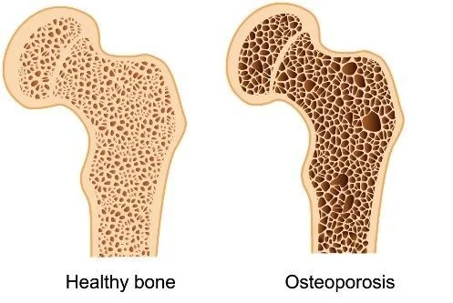
Osteoporosis is a disease that causes the bones to weaken, making them susceptible to breakage.

Osteoporosis occurs when the bones lose minerals quicker than the body can replace them, causing bone density loss. As bones become more porous, a minor fall, bump, sneeze or sudden movement may cause a fracture.

People may assume that Osteoporosis is associated with older women, however it can also affect men. One in three women over the age of 50 and one in five men over the age of 50 experience osteoporosis.

What causes it?

While the majority of our bone density is decided by genetics, lifestyle patterns during childhood and teenage years also play a role in your bone density.

By the age of 21, 95% of bone density is developed with the final 5% having developed by the age of 25. Your childhood and teenage years are important in building up your bone bank and increasing bone density. The higher the bone density during these years, the longer the onset of Osteoporosis later on in life. Eating a healthy diet with appropriate levels of calcium and exercising regularly can assist in building strong and healthy bones.

So, what can be done to minimise the effects of bone resorption?

Lifestyle choices play a huge role – especially exercise!

Weight bearing and resistance exercises can assist in bone reformation as well as strengthening the muscles to support the bones. It is important to strengthen the extensor muscles (back muscles), thoracic spine, and the muscles surrounding the shoulder blades. By strengthening these muscle groups, it can assist in developing postural strength/endurance which can reduce the chance of fractures.

Where does Pilates come in?

Pilates is an ideal form of exercise for those with Osteoporosis as well as for those looking for prevention. Pilates can:

  • Increase postural strength and support

  • Build and promote bone density

  • Correct posture

  • Improve alignment of the limbs

  • Reduce Osteoporosis symptoms

The exercises are provided by qualified Physiotherapists, with a specific focus on controlled loading of the bones.

So, come and see us in the UPG studio for an initial Pilates assessment and start putting some deposits in the bone bank today!

Written by Erin Gleadell: Pilates Lead and Physiotherapist - UPG Studio

References:


1. Angin, E., Erden, Z., & Can, F. (2015). The effects of clinical pilates exercises on bone mineral density, physical performance and quality of life of women with postmenopausal osteoporosis. Journal of Back and Musculoskeletal Rehabilitation, 28, 4, 849-858.

2. Rachner, T. D., Khosla, S., & Hofbauer, L. C. (2011). Osteoporosis: now and the future. Lancet (london, England), 377, 9773, 1276-87.

3. Tu, K. N., Lie, J. D., Wan, C. K. V., Cameron, M., Austel, A. G., Nguyen, J. K., Van, K., ... Hyun, D. (2018). Osteoporosis: A Review of Treatment Options. Pharmacy and Therapeutics, 43, 2, 92-104.

How Skipping Can Improve Your Running

Image Source: Pinterest

Image Source: Pinterest

Skipping is a form of exercise that can be highly beneficial for numerous reasons. New research has shown that skipping can be highly beneficial in improving your running performance.  The value of this simple form of exercise is often overlooked. A form of plyometric training, which involves rapid muscles contractions, as well as utilises the spring-like mechanism in our tendons. 

Running is an extremely popular form of exercise that reaps benefits for both our mental and physical wellbeing. Unfortunately, up to 80% of runners will experience a running related injury. This is often attributed to the high levels of load going through the joints of our lower limbs and the lack of adequate capacity our muscles have to withstand these forces. 

Load management is an integral consideration  for any runner, and it is important to gradually build up your running distance over time to avoid overloading, which can lead  to injury. In an effort to achieve running goals, runners can get injured by running too far and too soon. This is where skipping comes in as an alternative option to improve your running without having to simply increase your weekly kilometres.

How can skipping help you run better? For starters, studies have shown that skipping has less Ground Reaction Force on your knees, which are one of the more commonly injured joints in runners. Secondly, skipping has been shown to have a higher metabolic demand by up to 30% and more oxygen consumption. This means that you burn more calories and maintain your cardiovascular endurance required for running, all the while placing less force on your body. 

Research shows that skipping 2-4 times per week over a 10 week period for 10-20 minutes resulted in improvements in both running speed and improved times. Specifically, the study  showed significant improvements in performing 3km time trial running. 

Physiotherapists have the ability to assess the appropriateness of your running routine and can assist you in having a balanced exercise program, such as in implementing skipping. 


References: 
Garcia-Pinillos, et al, 2020
McDonnel et al, 2019

Why and How You Should Be Foam Rolling

IMG_1918.JPG

Looking after your health and well-being is important now more than ever as the COVID-19 lockdowns continue. Having to adapt to a different working environment and exercise routine can lead to your body feeling increased muscle tension, making it all the more important to practice extra self-care.

What is foam rolling, and why should I do it?

The foam roller has become a popular and convenient way to release muscle tension in the comfort of your own home. Not to mention it helps improve flexibility! Foam rolling is a method of self-massage that effectively reduces muscle tension and pain. When carried out before a workout, it can enhance physical performance and joint flexibility. Post-exercise, it can reduce the onset time and severity of muscle soreness, and minimise the associated acute muscle fatigue that impacts your power and speed. Moreover, doing foam rolling exercises at the end of the day can relax your body before going to bed. 

If you have access to one at home, then you are ready to start the session. Otherwise, there are different types available in varying shapes, sizes and textures on the market, which are affordable to purchase. The most popular and easiest to use is a high-density foam model. 

Our physiotherapist Anna Diep is going to guide you through some effective foam rolling techniques which will target the larger muscle groups in your body. With UPG Studio’s lead yoga instructor Keran Murphy demonstrating, we hope you benefit from this post.

How do I foam roll?

A few tips to consider before you start:

  • Complete each exercise for around 30-60 seconds (or slightly longer if you feel like it needs a bit more work).

  • Choose to incorporate both static stretching (keeping the roller still) as well as dynamic stretching (moving the roller over the muscle).

  • Ensure you’re rolling at a slow to medium speed to increase the effectiveness.

  • If you are new and still familiarising yourself with foam rolling, start with a lighter pressure and build up the pressure as you become familiar with the techniques. You should feel a moderate amount of discomfort through tender areas in the muscles but you should not be in excruciating pain. 

  • Ensure you keep breathing whilst foam rolling and avoid holding your breath. Taking a deep breath in slowly, holding for a couple seconds, then releasing slowly can help relax your muscles further for any of those extra tight spots.

  • Try to incorporate foam rolling into your regular routine (every 2 to 3 days).

Upper Back and Pectorals (Chest)

animated 3.GIF
  • Place the roller vertically in line with your upper spine.

  • Knees should be bent, with your feet pressed firmly onto the floor.

  • Lie on top of the roller, with your arms out relaxed beside you.

  • As your shoulders drop towards the floor, you should feel a stretch across your chest.

  • Slowly move your arms above your head and down beside your hips a few times to intensify the stretch.

Upper/Lower Back Roll

animated 2.GIF
  • Lay on your back with your knees bent, cradling your head in the palm of your hands with the foam roller across your shoulder blades.

  • Have your knees slightly bent. Lift your hips up off the floor and use your legs to roll your body up and down the foam roller. 

  • Aim to relax your upper body and let your spine flow gently backwards.

Quadriceps (Front of your Thigh)

animated 5.GIF
  • Start on your hands and knees with the roller underneath you.

  • Lay over the roller with either 1 leg (easier alternative) or both legs as demonstrated.

  • Walk your elbows forwards and backwards slowly, rolling between your hips and above your knees.

Gluteus Maximus (Bottom)

animated 7.GIF
  • Sit on the foam roller placing the one leg over the other at a 90 degree angle with your ankle above your knee.

  • Support yourself with the arm that is on the same side as the leg that is crossed, while gently pushing the knee down with the opposite arm.

  • Use the supported leg to gently push yourself forwards and backward in a range of 10-15cm, applying pressure to the whole outer portion of the outer hip. You can also rock from side to side or maintain a sustained pressure over any of those tight spots.

Hamstrings (Back of your legs)

animated.GIF
  • Sit on the foam roller with one leg extended up and the other leg bent for support, arms placed slightly behind you.

  • Using your arms and the supporting leg, shift your weight forwards and backwards so that the roller is moving between your bottom and your knees.

Iliotibial band (Side of your legs)

animated 2.GIF
  • Lie on your side with the foam roller underneath your hips, with your bottom forearm resting in line with the shoulder and your top arm in front of your chest.

  • Cross your top leg over to the front, while the bottom leg remains straight. Use your supporting leg and arms to guide the roller between your hips down to your knees.

Calves

animated 8.GIF
  • Start by sitting on your bottom with your arms placed slightly behind you.

  • Place both of your legs over the roller and lift your bottom off the floor.

  • (Easier alternative: Place one leg over the roller with the other leg bent. Push through your supporting leg and lift your bottom off the floor).

  • Use your arms to shift your weight forwards and backwards, with the roller moving between your ankles and below your knees.

If you are experiencing any injury or pain, our physios are here to help. Our South Yarra and Malvern clinics are still offering 1:1 physiotherapy consultations (with all necessary precautions in place), as we remain an essential health service. 

Book an appointment with us so that we can prescribe specific exercises and treatment to help you reduce symptoms, as well as provide advice on how to prevent future pain. 

If you are unable to attend a physiotherapy appointment in person, contact our South Yarra clinic at info@unitedphysiogroup.com.au to arrange a telehealth consultation. 

Stay safe, healthy, and check in on one another during this period. Happy foam rolling!

The Kapalabhati breathing technique explained.

Our yoga teacher Annabel practicing Kapalabhati.

Our yoga teacher Annabel practicing Kapalabhati.

Kapalabhati, also known as breath of fire, is a pranayama - a yogic breathing technique that cleanses, detoxifies and invigorates your mind and body. The word kapalabhati is made up of two Sanskrit words: kapāla meaning 'skull', and bhāti meaning 'shining, illuminating'.

A fiery technique, it best done before an asana flow, rather than after. It is also best practiced on an empty stomach, sitting in an upright position such as crossed legged or on the shins.

Our yoga teacher Annabel explains step by step how to practice kapalabhati, which she regularly starts her classes with.

  1. Start by inhaling fully, taking your time and filling up your lungs.

  2. Then, begin by exhaling shortly and sharply through the nose for anywhere from 50 to 120 strokes (exhales). Your focus should mostly be on the exhales, letting your inhales happen naturally. Passively inhale, actively exhale.

  3. When finished the strokes, exhale deeply, then inhale again, completely and hold your breath for a few counts.

  4. Finally, exhale completely and return to the normal rhythm of breath. 

Tips: When practicing, think about puffing the belly in and out, sucking the navel towards the spine on the exhale. It can also be beneficial to place a block under your sit bones in order to align your spine, and to place your hands on your lower belly. 

Some other benefits include:

  • An increased amount of prana (energy) moving throughout your body. Notice how your body feels after the practice.. you might feel more energised, as an increased amount of blood will now be circulating throughout the your body. 

  • Kapalabhati also helps to calm the mind. When we are so focused on the ratio, pattern and force of the breath, we have no time to think of anything else. This allows our mind to settle and focus. 

If you are looking to practice yoga, South Yarra’s UPG Studio offers intimate, personalised classes to help you get the most out of your practice. Book a class with us here.

Yoga 'Pigeon' Pose

The ‘Pigeon’

Benefits of Half Pigeon include:

  • Release and opening of the hips, a place where we often hold a lot of built up emotions, especially for women. I find that it is beneficial for me to release stagnant energy and anxieties which I often hold in my hips

  • Stretches quads and glutes

Half pigeon pose is fairly accessible to all as it can be modified. Great options if you need a bit more support in pigeon include:

  • A block under the hip

  • A block under the forehead or arms

  • Remaining upright in seated half pigeon rather than folding all the way in

If you'd like to deepen the pose, some good options include:

  • Folding in over the top leg onto forearms or all the way to the floor

  • Moving into full mermaid by sitting upright and bending into the knee of the back leg, grabbing hold of your back foot. Option to bind here by bringing the elbow crease to the foot and interlocking the hands

  • Moving into full pigeon by swinging the back leg around and stacking it on top of the lower leg, with both shins parallel with the top of the mat

If half pigeon is not accessible to you a great alternative is reclined figure 4. To get into this shape (on the right side):

  • Lay on your back

  • Plant both feet flat on the floor with knees bent

  • Lift your right leg and place the ankle on top of the left knee, opening the right knee out to the side

  • Then grab hold of the back or front of your left knee and pull in towards your chest as much as feels comfortable

IMG_6124.JPG
My favourite thing about yoga is: That it is for everyone.
— Annabel Macfarlane
IMG_6123.JPG